ROTATOR CUFF/INPIGIMENT
The shoulder is not a single joint, but a complex of 4 articulations, several tendons, and many muscles that interact to create the most mobile “joint” in the human body. With many activities, including rigorous athletics, there is an appreciable demand on the shoulder. Injury resulting in pain and loss of function occurs when the physiologic limits of these tissues are exceeded.
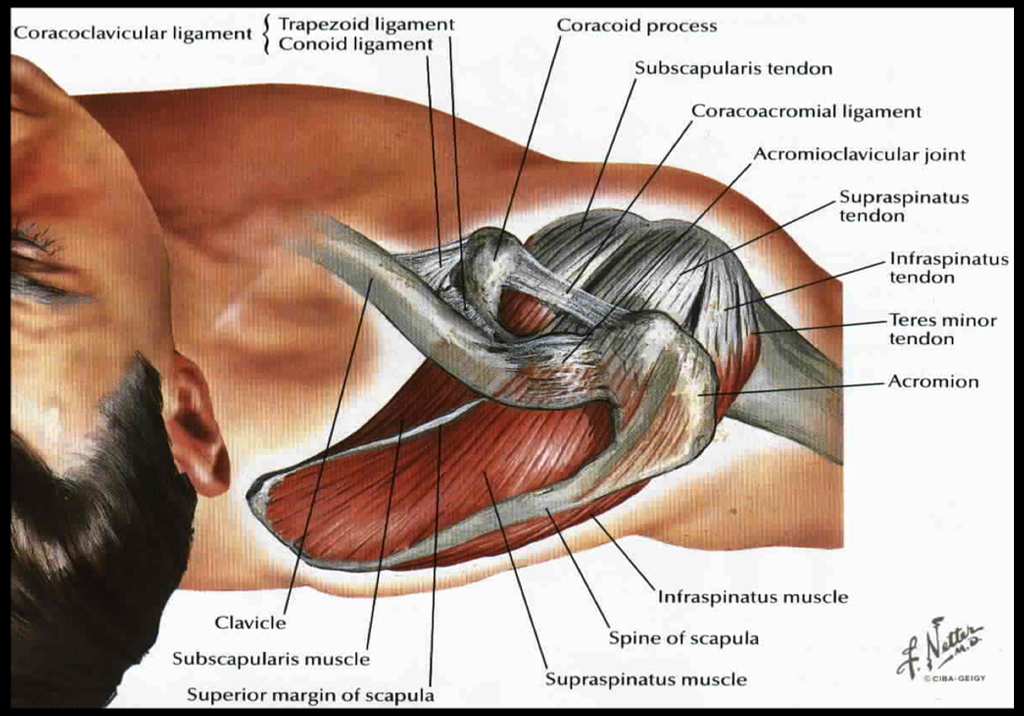
These articulations include the scapulothoracic articulation, or where the shoulder blade runs on the posterior chest wall, the sternoclavicular joint, or where the collar bone(clavicle) meets the sternum(breast bone), the acromioclavicular joint (AC joint) where the clavicle meets the acromion bone, and the glenohumeral joint, the main shoulder joint most commonly associated with the term “shoulder”. Injury to any of these articulations singly or in combination can cause pain and loss of shoulder function.  Four muscles comprise the rotator cuff : the supraspinatus, infraspinatus, subscapularis, and the teres minor. Of these, the first three are the most commonly involved in rotator cuff disease. The supraspinatus, infraspinatus, and teres minor join to form a common tendon that connects these muscles with the humerus bone at the shoulder. The rotator cuff forms the “roof” of the glenohumeral joint. The subscapularis tendon is separate from the remainder of the cuff, sitting in front of the glenohumeral joint, and separated from it by the biceps tendon. The function of the rotator cuff is to keep the humeral head (ball) centered on the glenoid (socket) during elevation of the arm, while the bigger muscles such as the deltoid, pectoralis, latissimus and trapezius provide the power. If the cuff doesn’t function properly, either from tearing or from weakness , the humerus bone will displace upward and pinch the cuff against the acromion bone (the bone on “top” of the shoulder where the Deltoid muscle attaches), causing “impingement” and resultant pain. The rotator cuff functions as the transmission of the shoulder. If the muscles are the engine, the rotator cuff is the linkage between motor(muscles) and the wheels(arm). If the motor is great, but the transmission is broken, the car wont work right. It’s the same with the shoulder. The rotator cuff runs beneath the acromion and can also be impinged by spurs on its undersurface. A Bursa normally sits between the rotator cuff and the acromion bone. This can become quite inflamed with an impingement syndrome and cause pain. The subscapularis is the anterior component of the cuff and functions to internally rotate the and flex the humerus. It also provides stability to the glenohumeral joint.
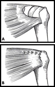
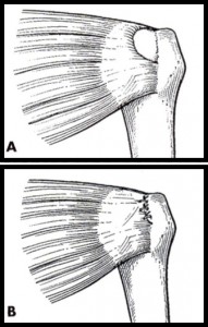
Repetitive activity, overuse, and falls are common causes of rotator cuff dysfunction. Almost all patients with cuff tears demonstrate degeneration of the cuff itself and sometimes minor events may cause tearing in this weakened tissue. Most shoulder problems can be managed successfully with therapy aimed at restoring normal cuff strength and stretching to reduce impingement type symptoms. Home programs initially usually are not sufficient and formal therapy with instruction is preferred. Cortisone shots can provide short term pain relief, but multiple injections may cause weakening of the rotator cuff and have been implicated in tearing of the cuff. If after several months of conservative treatment no significant improvement is seen, arthroscopic decompression may be curative. If an mri shows a cuff tear, however, a repair is likely necessary to fix the problem.
If a torn cuff is suspected on presentation, an mri will be done earlier. A torn rotator cuff almost never heals on its own, and like other torn items (fabric, clothing, etc,), the neglected tear tends to enlarge with time. This concept of timely rotator cuff repair is important because the tears tend to enlarge silently in 40% of patients. Orthopedic studies have shown far superior outcomes in terms of function and cuff healing in tears that are repaired early before fatty infiltration and atrophy of the muscle occurs. These changes are irreversible, and once initiated tend to progress even after successful repair has been done. As common sense would dictate, small tears are more easily repaired and have better outcomes than large tears, and the orthopedic literature confirms this.
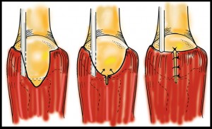
The surgery is done as an outpatient procedure with the use of general anesthesia combined with a nerve block to help with pain control. I do only arthroscopic repairs and all tears, regardless of the size , can be managed without opening the shoulder. Because the deltoid muscle isn’t detached with the arthroscopic approach, there is less pain postoperatively. A sling must be worn for 6 weeks, sometimes longer in massive tears, to protect the repair. Too vigorous activity in the early postoperative period can cause failure of the repair. Most commonly this occurs because the degeneration in the cuff tissue weakens it to such an extent that the stitches can cut through it. Physical therapy is necessary as well. Studies have shown improved motion after arthroscopic cuff repair if formal therapy is instituted at 4-6 after surgery, rather than earlier. This seems counterintuitive but has been borne out in several elegant studies. Strengthening begins at 3 mos in most patients and return to sports in 4-6 months depending on the demands of the activity.


